Regular events
Throughout the year, we have weekly CMCB lab talks. Click here for details of the current programme, format, and rules.
There is a weekly Cake Club, for aspiring bakers in CMCB. Details here or follow along on Twitter.
We have a monthly happy hour organised by a different research groups on a rota. Last Friday in the month, 1630 in the CTU Atrium or outside by the pond in sunny weather(!).
You can stay up to date with CMCB events and activity by following us on Mastodon. To read an archive of what happened in the past, click here.

Our next event
We are organising Motors in Quarantine Talks. Expressions of interest and further info here

Archive of past events
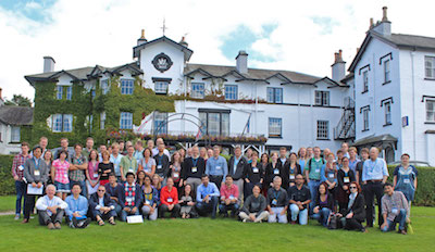
We have organised events, meetings and more in the past. Here is an archive of the pages:
- Mechanobiology at The Shard 2018
- Mechanobiology at The Shard 2017
- The Dynamic Cell II, 2014
- Mechanochemical Cell Biology, 2013
- UK-Japan, 2012
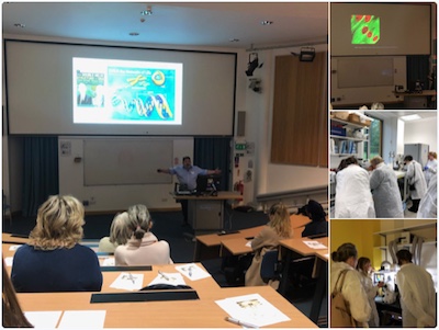
We also took part in New Scientist Live, ExCeL London 20-23 Sep 2018.